News Center
Innovative Solution Provider for Minimally Invasive Surgery

Column Introduction:
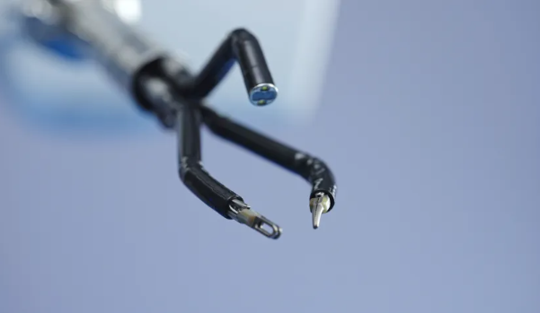
"Edge Medical Robotics Clinical Application Notes" is a professional content column focusing on real-world clinical practice of Edge surgical robotics. Taking clinical needs as the starting point and authentic surgical procedures as the vehicle, it systematically presents key surgical steps, instrument coordination logic, technical application value, and surgeon experience regarding the application of Edge Medical robotic across different specialties, different procedures, and complex clinical scenarios. Through each surgery and every operative detail, we aim to document the process of deep integration between robotic technology and clinical needs, providing more referable practical evidence for the refinement, standardization, and innovative development of minimally invasive surgery.
Prostate cancer is one of the most common malignancies in elderly men. For selected patients with localized prostate cancer, radical prostatectomy remains an important treatment modality. With the continuous advancement of surgical techniques, radical prostatectomy has progressively evolved from open surgery and laparoscopy into the era of robot-assisted surgery.
While technology evolves, the goals pursued by surgeons remain clear: to achieve oncological radicality while preserving critical anatomical structures related to urinary continence and sexual function, and to balance postoperative functional recovery with quality of life.
Guided by this objective, the Urology Department of the First Medical Center of Chinese PLA General Hospital, led by Academician Zhang Xu, has long been engaged in technological innovation in robotic prostate cancer surgery. Building upon the conventional anterior approach, the team has developed a technical system of robot-assisted radical prostatectomy with Anterior Preservation of Pelvic Floor Structures (APSS) through a series of technical optimizations including anatomical preservation of the bladder neck, protection of key pelvic floor structures, and functional reconstruction.
Recently, Professor Wang Baojun from the Urology Department of the First Medical Center of Chinese PLA General Hospital performed extraperitoneal robot-assisted radical prostatectomy with preservation of pelvic floor structures (SP-RARP) using the Edge Medical® Single-Port Endoscopic Surgical Robot (SP1000 Plus). Within the narrow extraperitoneal space, the team leveraged the single-port robotic visual field and instrument coordination to complete anatomical preservation of the bladder neck, total intrafascial dissection of the prostate, neurovascular bundle preservation, functional urethral preservation, and vesicourethral anastomosis with pelvic floor reconstruction, further exploring the integrated application of APSS technique and the single-port robotic platform.
Extraperitoneal Approach
Achieving Fine Dissection and Structural Preservation within a Limited Space
The patient was a 74-year-old male diagnosed with prostatic acinar adenocarcinoma by prostate biopsy. Based on Gleason score, biochemical, and imaging findings, the case was assessed as intermediate-risk localized prostate cancer.
Considering the patient's age, pulmonary function, and other factors, the surgical team selected the extraperitoneal approach for robot-assisted radical prostatectomy after comprehensive evaluation.
Following anesthesia, a approximately 3 cm main operative incision was made below the umbilicus. An extraperitoneal operative space was established between the posterior rectus sheath and the peritoneum, and an additional assistant port was placed. The Edge Medical® Single-Port Endoscopic Surgical Robot was then docked, and the endoscope and SuperArm surgical instruments were installed.
Unlike the transperitoneal route, the extraperitoneal approach directly enters the retropubic space, where the operative space is relatively limited. For the APSS technique, the surgeon is required to perform fine dissection around critical structures including the bladder neck, endopelvic fascia, dorsal venous complex (DVC), neurovascular bundles, prostatic apex, and functional urethra, placing high demands on surgical field exposure, instrument maneuverability, and deep suturing capability.
The procedure lasted approximately 75 minutes, and all planned surgical steps were completed successfully.
SuperArm Instrument Formation
Organized around "Cutting, Dissecting, Retracting, and Suturing" Tissue Operations
Monopolar Scissor
Sharp dissection | Flexible articulation | Precise transection
SuperArm Monopolar Scissors: The distal end features a scissor structure that allows flexible adjustment of the cutting angle within deep and narrow spaces. The surgeon may select cold scissor sharp dissection or combine with monopolar energy for tissue cutting and management according to specific operative needs.
Interpretation: In this case, after the anatomical plane was clearly defined, the monopolar scissors were primarily used for bladder neck incision, sharp dissection of fascia and tissue, and management of the prostatic apex. The surgeon could choose cold scissor or monopolar energy-assisted dissection based on the specific anatomical situation. Particularly during the management of the prostatic apex and functional urethra, fine sharp dissection helped the surgeon complete tissue transection along the predetermined anatomical plane.
Bipolar Maryland
Traction and exposure | Fine dissection | Immediate hemostasis
SuperArm Bipolar Maryland: The slender jaw design enables tissue grasping, lifting, pushing, and blunt dissection within narrow spaces. Leveraging the multi-degree-of-freedom mobility of the robotic wrist, the surgeon can flexibly adjust the jaw orientation to establish a more suitable traction angle and dissection path.
Interpretation: In this case, in fine dissection areas including the bladder neck, Denonvilliers' fascia, and around the neurovascular bundles, the surgeon gradually established planes through traction and blunt probing, forming a continuous operative sequence of "exposure—identification—dissection—transection" in coordination with the monopolar scissors.
Needle Driver
Stable needle holding | Flexible articulation | Fine suturing
SuperArm Needle Driver: Used for surgical needle grasping, tissue suturing, and knot tying. Leveraging the multi-degree-of-freedom mobility of the robotic wrist, it allows flexible adjustment of needle-holding angle and needle insertion direction within deep and narrow spaces, making it suitable for anastomosis, reconstruction, and complex tissue suturing.
Interpretation: In this case, after completion of prostatectomy, the procedure transitioned from "resection" to "reconstruction." The surgeon exchanged for the needle driver and completed vesicourethral anastomosis within the deep pelvis, performing VPM and anterior structure reconstruction as appropriate based on intraoperative preservation status, carrying the concept of functional protection through to the final step of the surgery.
Key Step One:
Advancing along Anatomical Planes
Maximizing Preservation of Key Pelvic Floor Structures
After the robot entered the extraperitoneal space, the surgeon first pushed aside the anterior vesical fat, cleared the retropubic fat, and adequately exposed the bladder and anterior prostate.
Unlike some maneuvers in the conventional anterior approach, this case followed the APSS technique principles: the endopelvic fascia was not opened, and the dorsal venous complex (DVC) was not pre-ligated, minimizing disturbance to the normal periprostatic anatomical structures.
Subsequently, the assistant helped identify the bladder neck location by traction on the urinary catheter. The surgeon sharply incised the vesicoprostatic junction using monopolar scissors and, in coordination with bipolar dissecting forceps, identified the relatively avascular anatomical plane between the bladder musculature and the prostatic capsule, gradually completing bladder neck dissection while preserving structures such as the longitudinal muscle group (VPM) posterior to the bladder neck.
Continuing posterior dissection, the team completed mobilization of the vas deferens and seminal vesicles, and advanced along the plane between the prostatic capsule and Denonvilliers' fascia toward the prostatic apex, performing neurovascular bundle preservation when oncologically appropriate for the patient.
Throughout the procedure, the monopolar scissors were responsible for precise sharp dissection, while the bipolar dissecting forceps handled tissue traction, plane probing, and local hemostasis. The two instruments continuously coordinated around the principles of "clearly visualizing the plane, dissecting along the plane, and minimizing unnecessary structural disturbance."
Key Step Two:
From Functional Urethral Preservation to Pelvic Floor Reconstruction
Carrying "Protection" Throughout
The prostatic apex is a critical anatomical region in radical prostatectomy and one of the key steps in balancing oncological control with preservation of continence-related structures.
After completing periprostatic mobilization, the surgeon continued fine management of the detrusor apron, DVC, and anterior fibromuscular stroma (AFS), circumferentially mobilized the apical urethra, preserved functional urethral length as much as oncologically permissible, and completed urethral transection flush with the prostatic apex, achieving complete removal of the prostate.
The procedure then entered the reconstruction phase. The surgeon exchanged for the needle driver and completed vesicourethral anastomosis using continuous suturing, with corresponding reconstruction based on VPM preservation status; subsequently, anatomical reduction of the periurethral pelvic floor structures was further performed.
From the early phase of "no opening, no pre-ligation," to the middle phase of "dissecting along planes, preserving whenever possible," to the final phase of "fine anastomosis, reconstruction when necessary," the APSS technique is not a single surgical maneuver but a comprehensive anatomical preservation philosophy that spans the entire process of resection and reconstruction.
For the single-port robot, the real question is not simply "whether the surgery can be completed," but whether it can provide sustained, stable conditions for this series of fine operations within the limited extraperitoneal space.
Surgeon Experience (Professor Wang Baojun, Chinese PLA General Hospital):
Smaller Space Demands Greater Anatomical Understanding and Organization of Every Movement
Professor Wang Baojun stated that the operative space for extraperitoneal radical prostatectomy is relatively limited, and the APSS technique emphasizes fine identification, preservation, and reconstruction of key pelvic floor structures, thus placing high demands on the surgeon's anatomical understanding and instrument coordination.
After single-port robotic instruments enter the body through the same main operative channel, operative angles can be reorganized around the target area. In this case, the team performed targeted combinations among monopolar scissors, bipolar dissecting forceps, bipolar grasping forceps, and needle holder according to the tasks of different phases: the dissection phase emphasized coordination between traction and sharp dissection, the apical management phase emphasized tissue identification and cold scissor operation, and the reconstruction phase focused on needle-holding direction and suturing path within the deep pelvis.
For the surgeon, instruments are merely tools for technical realization. What truly determines surgical quality remains the accurate judgment of anatomical planes and the choices made for every movement around the goals of oncological control and functional preservation.
Summary:
The development of robot-assisted radical prostatectomy is driving prostate cancer surgery beyond a sole focus on "how to achieve complete resection" toward "how to more finely preserve and reconstruct function-related structures while achieving oncological goals."
The APSS technique proposed and continuously explored by Academician Zhang Xu's team is precisely one clinical practice under this philosophy.
In this case, Professor Wang Baojun's team combined the extraperitoneal APSS technique with the Edge Medical® Single-Port Endoscopic Surgical Robot, completing prostatectomy and complex pelvic floor structure preservation and reconstruction through an approximately 3 cm main operative incision, accumulating new clinical practice experience for the application of single-port robotics in refined urological procedures.
From the preservation of a single fascia, to the fine management of a segment of functional urethra; from a single anastomotic stitch spacing, to the reconstruction of a set of pelvic floor structures—when robotic technology truly enters the depths of clinical practice, its ultimate foundation remains the surgeon's understanding of anatomy, judgment of operative maneuvers, and unwavering pursuit of functional preservation for patients.
Contributor for this issue: Qi Hengzhi
(This article is intended solely for the sharing of clinical technique and instrument application experience. The content is based on the actual circumstances of this case and the surgeon's operative experience, and does not constitute universal recommendations for specific treatment plans or instrument selection.)

